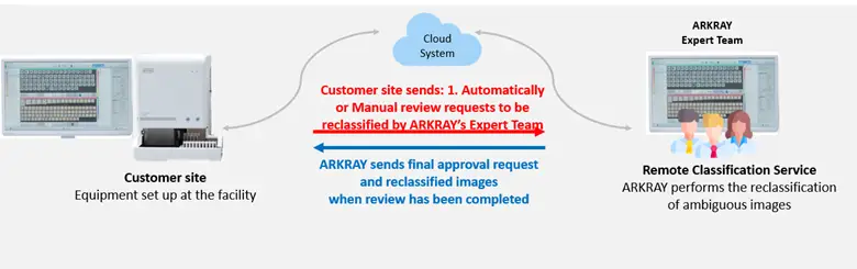
ARKRAY USA, Inc. Image Analysis supports image review and reclassification of automated classification results from urine formed element analyzers*, helping reduce microscopy rates and allowing laboratories to focus on samples that require detailed analysis. This contributes to greater efficiency and higher quality laboratory operations.
*This service requires the use of the AUTION EYE AI-4510 Analyzer.
Supporting Human Resource Challenges in the Laboratory
I have difficulty recruiting talent.
With AUTION EYE Smart Assist, clinical laboratory technologists experienced in urine sediment analysis are available to support your testing workflow. You can receive expert assistance in image evaluation without the time, effort, and cost associated with recruitment or training.
Sudden staffing shortages due to maternity leave or sudden absences
AUTION EYE Smart Assist offers flexible scalability to accommodate sudden changes in staffing. You can easily increase usage during months when additional support is needed, reducing the burden of overtime work and emergency staffing, while maintaining stable and efficient laboratory operations.
Limited time to train new staff.
The AI-4510 automatically stores test results as images, allowing staff to review formed element images at any time. In addition, results from the Image Analysis Center, including reclassification outcomes and atlas references can be accessed to support standardization of interpretations and serve as effective training material, eliminating the need to create separate educational tools.
Save time spent on manual microscopy
AUTION EYE Smart Assist helps significantly reduce the number of specimens that require microscopic examination. Images captured by the AI-4510 are reclassified at the Image Analysis Center to precisely narrow down samples that truly need manual review. This helps maintain analytical quality while improving overall laboratory efficiency.
Service Features
Managed by Certified Technologists and Quality Supervisors
ARKRAY USA, Inc. Image Classification Center is operated under the supervision of certified laboratory technologists who also serve as quality control managers. Leveraging extensive experience in urinalysis and general laboratory testing, they oversee system operations, staff training, and process management to ensure the highest standards of analytical quality.
Manual, Computer-Based Review by Dedicated Technologists
All image classifications are carefully reviewed by technologists on a computer with extensive experience in urine sediment testing. Each set of test data sent to the Image Analysis Center is carefully reviewed and, if necessary, reclassified. Technologists also verify whether any components requiring further microscopic examination are present, ensuring thorough and accurate feedback.
Secure, Guideline-Compliant Cloud Infrastructure
Our service platform is designed to meet U.S. standards for medical data security and privacy, ensuring reliable and compliant operation within clinical environments. It aligns with major regulatory frameworks, including:
The Health Insurance Portability and Accountability Act (HIPAA) for the protection of patient health information
The Health Information Technology for Economic and Clinical Health (HITECH) Act for secure electronic health data management
FDA and CLIA guidance for laboratory data integrity and system validation
This ensures a robust, secure environment for data handling, storage, and system reliability.
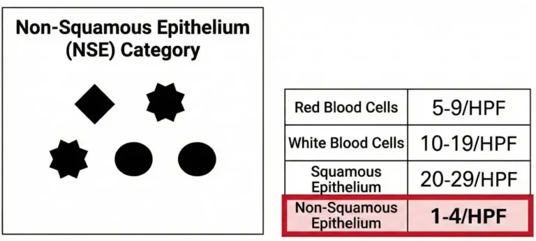
Reducing Microscopy Rates Through Image Reclassification
Before reclassification
When “non-squamous epithelial cells ≥1–4/HPF” are set as criteria for microscopic review, specimens automatically classified as non-squamous epithelium by AUTION EYE become targets for manual microscopy.
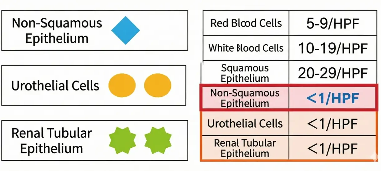
After reclassification
Through detailed reclassification at the Image Analysis Center, components initially labeled as non-squamous epithelium can be more precisely identified as urothelial or tubular epithelial cells. As a result, the number of specimens requiring microscopy decreases, reducing unnecessary manual review while maintaining diagnostic accuracy.